Advanced Diagnostic Testing for Ocular Diseases

Macular Pigment Testing

Macular pigment testing is a specialized procedure used to measure the density of the macular pigment in the eye. This pigment, located in the macula—the central part of the retina—is crucial for protecting the eyes against harmful blue light and maintaining sharp, detailed central vision. Low macular pigment density can be a risk factor for age-related macular degeneration (AMD).

The test is simple, non-invasive, and provides valuable information about the health of the macula. It involves looking into a device that measures the eye’s response to different light stimuli. Knowing the density of the macular pigment can help in recommending dietary or lifestyle changes, like increasing intake of lutein and zeaxanthin, which are known to enhance macular health.

Comprehensive Visual Field Testing

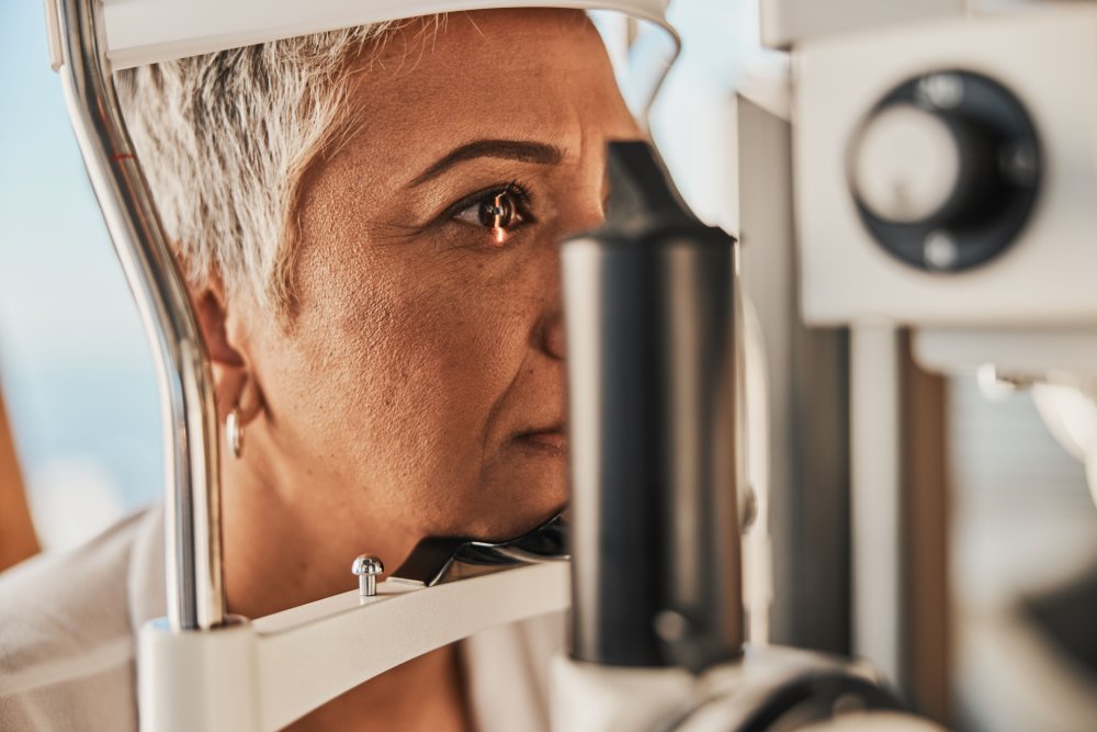
Comprehensive visual field testing is a crucial part of eye exams, especially for diagnosing and managing conditions like glaucoma. This test measures the entire scope of your vision, including peripheral vision, to detect any blind spots or vision loss. Visual field testing is essential in identifying early stages of glaucoma, where peripheral vision is often affected first.

The test involves looking at a screen and indicating when a light is seen in the peripheral vision. This helps map out any areas where vision may be compromised. Regular visual field testing is important for patients at risk of glaucoma, as it helps in monitoring the disease’s progression and the effectiveness of the treatment.

Advanced diagnostic testing is crucial in the early detection and management of various ocular diseases. These tests use state-of-the-art technology to provide detailed images and information about the structures within the eye. They can detect conditions like glaucoma, diabetic retinopathy, and age-related macular degeneration at an early stage when they are most treatable.

Some common advanced diagnostic tests include optical coherence tomography (OCT), visual field testing, and digital fundus photography. Each of these tests provides essential information about different aspects of the eye, allowing for a comprehensive evaluation of ocular health and aiding in the development of an effective treatment plan.

Fundus Photography

Fundus photography is a diagnostic procedure that captures detailed images of the retina, the optic disc, and the blood vessels at the back of the eye. This test is critical in diagnosing and monitoring retinal diseases like diabetic retinopathy, age-related macular degeneration, and retinal detachment. It provides a baseline to track changes over time and can help in early disease detection.

The process is non-invasive and quick, involving the use of a specialized camera to photograph the fundus. These high-resolution images allow optometrists to detect and assess any abnormalities or changes, facilitating early intervention and management of ocular conditions.

Ocular Coherence Tomography (OCT)

Optical Coherence Tomography (OCT) is a non-invasive imaging test that uses light waves to take cross-sectional pictures of your retina. OCT provides detailed images that can show the different layers of the retina, allowing for the detection of conditions such as glaucoma, macular holes, and changes related to diabetes.

The OCT scan is particularly important in the early detection and management of macular diseases, including macular degeneration and diabetic eye disease. The detailed view it provides helps in diagnosing subtle changes that might not be visible through a regular eye exam. This tool is essential in tracking the progression of retinal diseases and assessing the response to treatment.

Zeiss Humphrey Visual Fields

Zeiss Humphrey Visual Field Analyzer is a sophisticated tool used for visual field testing. It’s particularly effective in detecting, diagnosing, and managing glaucoma, as well as other neurological conditions that affect vision. This advanced system provides detailed and reliable data, making it a gold standard in visual field testing.

The Humphrey Field Analyzer uses automated perimetry to map the visual field, detecting any areas of vision loss

Heru Portable Visual Fields

The Heru Portable Visual Fields test represents a modern advancement in visual field testing, offering a more flexible and convenient approach compared to traditional methods. This portable device can perform visual field tests without the need for a large, stationary machine. It’s particularly useful for patients who have mobility issues or in settings where space and resources are limited.

Utilizing virtual reality technology, the Heru device can accurately assess a patient’s visual field, detecting any defects or blind spots. This is crucial for diagnosing and monitoring conditions like glaucoma, where peripheral vision loss is a key indicator. The portability and ease of use make it an ideal choice for regular monitoring, especially in community health settings or for home visits.

ICare Tonometer

The ICare Tonometer is an innovative device used for measuring intraocular pressure (IOP), a key factor in glaucoma management. This device offers a quick, painless, and accurate way to measure eye pressure without the need for numbing drops, making it more comfortable for patients.

Unlike traditional methods, the ICare Tonometer uses a tiny probe to gently make contact with the eye’s surface, requiring minimal cooperation from the patient. This makes it particularly useful for children, elderly patients, or those who are sensitive to the air puff test. Regular IOP monitoring is essential in diagnosing and managing glaucoma, and the ICare Tonometer provides a patient-friendly option for this important test.

Request Appointment